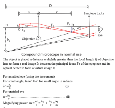
Compound Microscope Normal Adjustment
Result and Interpretation of Gram Staining. Use the coarse adjustment knob to get a general focus.
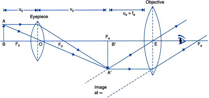
Draw A Ray Diagram Of A Compound Microscope Write The Expression For Its Magnifying Power From Physics Ray Optics And Optical Instruments Class 12 Cbse
Compound eyes are very.
. Microscope with 100X objective lens compound microscope. The Journal of Prosthetic Dentistry is the leading professional journal devoted exclusively to prosthetic and restorative dentistryThe Journal is the official publication for 24 leading US. However stereo microscopes offer lower magnification typically 5x-50x comparing to compound microscopes.
The monthly publication features timely original peer-reviewed articles on the newest techniques dental materials and research findings. It comes with a sliding binocular viewing head two pairs of widefield eyepieces WF10X WF20X four achromatic objectives DIN 4X 10X 40XS 100XS Oil large double layer mechanical stage with scale Abbe NA125 condenser with iris diaphragm coaxial coarse. Li et al 2021.
Center the slide so that the specimen is under the objective lens. This is the maximum Basic English combined wordlist. Gram-Negative bacteria appear pink or red.
The microscope stage should be lowered as low as possible. The main objective of the present study is to evaluate the efficacy of foldscope and also used to compare foldscope and normal light microscope. PDF On Jan 1 2014 Naveena Varghese and others published Microbiology Laboratory Manual Find read and cite all the research you need on ResearchGate.
OMAX compound biological microscope comes with eight level magnifications from 40X to 2000X. This diagnostic tool is helpful in learning biology. The basic differential interference contrast DIC system first devised by Francis Smith in 1955 is a modified polarized light microscope with two Wollaston prisms added one to the front focal plane of the condenser and a second at the rear focal plane of the objective see Figure 1Several years later Georges Nomarski a Polish-born French physicist modified the.
Start with a low lens and a clean slide. Unlike a compound microscope that can only see a very thin specimen stereo microscopes can be used for viewing almost anything you can fit under them. 5 mL of sample was digested with an equal volume of simulated gastric fluids SGF 2 mgmL NaCl 32 mgmL pepsin pH 2 at 37 C 200 rpm for 2 h followed by immediate pH adjustment to neutral and addition of 10 mL of.
In 50 of cases the psychiatric symptoms appear first. Gram-positive cocci Staphylococcus spp. Gram-Positive bacteria appear violet or purple.
How to Use a Light Microscope. It is what the advanced student will know when moving from Basic English to the standard English language. The compound eyes of the arthropods are composed of many simple facets which depending on anatomical detail may give either a single pixelated image or multiple images per eye.
Some eyes have up to 28000 such sensors arranged hexagonally which can give a full 360 field of vision. Then slowly move up the. Signs and symptoms of Huntingtons disease most commonly become noticeable between the ages of 30 and 50 years but they can begin at any age and present as a triad of motor cognitive and psychiatric symptoms.
The difference between Compound and Stereo Dissecting Microscope. Below is an. Each sensor has its own lens and photosensitive cells.
The simulated digestion experience was carried according to previous work with some modifications Chen Zhao et al 2021. To use a light microscope you can follow the steps below carefully. Focus the microscope using a fine adjustment knob and study the bacteria.
At the top of our list youll find the AmScope 120X-1200X 52-pcs Kids Beginner Microscope STEM Kit M30-ABS-KT2-W. Examples of Gram-positive bacteria. The polyphenol procyanidin C1 a compound found in grape seeds possesses senomorphic or senolytic activity and is shown to extend the healthspan and survival of old mice and in various models of.
Their progression is often described in early stages middle stages and late. This powerful compound microscope for kids is more than just a tool its a.
Schoolphysics Welcome
Draw A Neat Labeled Ray Diagram Of A Compound Microscope Explain Briefly Its Working Sarthaks Econnect Largest Online Education Community

Draw A Labelled Ray Diagram Showing The Formation Of Image By A Compound Microscope In Normal Adjustment Derive The Expression For Its Magnifying Power Physics Shaalaa Com
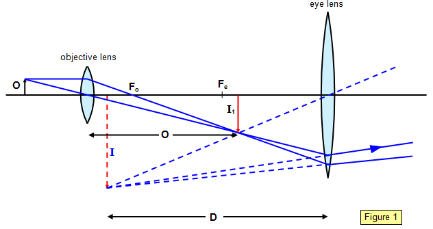
Draw A Diagram To Show The Formation Of An Image By A Compound Microscope In In Normal Adjustment And Use It To Derive An Expression For Magnifying Power Digital Teachers Uganda


Comments
Post a Comment